TL;DR — Visual Aids for Dental Patient Education
- Visual aids increase patient comprehension of dental diagnoses by 40–60% compared to verbal explanation alone — and comprehension drives treatment acceptance.
- Intraoral cameras are the highest-ROI visual tool in dentistry: they show patients their own mouth, eliminating the credibility gap that slows treatment acceptance.
- 3D animation and digital models improve case acceptance rates for complex cases (implants, full-arch restoration) by 25–35%.
- Before-and-after photography builds case acceptance for cosmetic cases and drives organic social proof — two outcomes from the same 30-second action.
- Visual education works best when it’s integrated into the appointment flow, not bolted on as an add-on presentation.
The most powerful treatment presentation tool in a dental practice isn’t a brochure, a verbal explanation, or a fee quote — it’s a clear image that shows the patient exactly what you see. When patients can see their own cavity, their receding gumline, or the fracture running down their molar on a screen in front of them, the credibility gap between “the dentist says I need this” and “I can see why I need this” disappears. That gap is where treatment acceptance is lost.
For related reading, see our guide on growing through patient retention.
This guide covers the visual tools that produce the best clinical communication outcomes — with specific data on which tools move case acceptance rates, how to integrate them without lengthening appointments, and the implementation sequence that produces the fastest measurable results. The education component of patient relationships fits within the broader framework of our pillar guide on building patient trust and relationships.
For related reading, see our guide on reducing patient no-shows.
Why Do Visual Aids Improve Dental Patient Education and Case Acceptance?
The cognitive science underlying visual dental education is well-established. Humans process images approximately 60,000 times faster than text, and visual information is retained at significantly higher rates than verbal information alone. A 2019 study in the Journal of Dental Education found that patients who received visual explanations of their dental diagnoses demonstrated 58% better recall of the recommended treatment plan one week later than those who received verbal explanations only — and were 32% more likely to have scheduled the recommended treatment (JDE, 2019).
In the dental context, visual aids serve three distinct functions:
- Diagnosis validation: Showing patients evidence of a problem removes the “but it doesn’t hurt” objection that delays treatment for asymptomatic conditions.
- Treatment comprehension: Explaining a crown prep or implant placement with a 3D animation is categorically more effective than verbal description for patients without clinical training.
- Emotional engagement: Before-and-after photography and outcome simulations connect patients to the result they want, engaging motivation that clinical necessity alone doesn’t always activate.
What Are the Most Effective Visual Tools for Dental Patient Education?
Intraoral Cameras
The intraoral camera is the single highest-impact visual tool for case acceptance in general dentistry. When a patient can see their fractured cusp, their failing restoration, or their periapical pathology on a chairside screen in real time, treatment recommendation transforms from the dentist’s opinion to shared clinical evidence. Practices consistently report case acceptance rate improvements of 20–40% on restorative cases after implementing intraoral cameras with a consistent chairside presentation protocol.
Implementation best practices: the image should be displayed on a monitor the patient can see without turning their head, the dentist should describe what the image shows in plain language while pointing to the specific finding, and the image should be saved to the patient record for use in follow-up communication. Images saved to the patient record and referenced at future visits (“Here’s what we saw last November — notice how this has changed”) create a powerful visual timeline that motivates treatment urgency.
Digital Radiography With Patient-Facing Displays
Digital X-rays have replaced film in most practices, but many practices still review radiographs on a monitor the patient cannot easily see. Repositioning the display — or using a secondary patient-facing monitor — to include the patient in radiograph review transforms X-rays from a diagnostic tool used by the dentist into an educational tool shared with the patient. Simple annotations (circling the area of concern, adding a brief text label) make the clinical finding accessible to a lay patient immediately.
3D Dental Animations
For complex cases — implants, full-arch rehabilitation, periodontal surgery, root canals — animated explanations of the procedure dramatically improve patient comprehension over verbal description. Platforms like Dental Patient Education (DPE), Caesy, and Guru provide libraries of professional-quality dental animations organized by procedure type. A 2021 review in BMC Oral Health found that patients who received video/animation-based treatment explanations demonstrated 35% better procedural comprehension and reported lower pre-treatment anxiety than patients who received verbal explanations alone (BMC Oral Health, 2021).
Integration tip: the most effective workflow is not to show an animation instead of a verbal explanation, but to show a 60–90 second animation first, then discuss. The animation primes the patient’s understanding; the conversation that follows is more productive because the patient has a mental model of what you’re describing.
Before-and-After Photography
For cosmetic and elective cases — veneers, bonding, whitening, clear aligners — before-and-after photography is the most persuasive case acceptance tool available. Outcome photographs work because they allow patients to envision their own result by proxy. The key is maintaining a well-organized photo library that includes cases similar to the patient’s specific situation: age range, starting condition, and desired outcome.
Before-and-after photos also generate organic social proof when shared (with patient consent) on the practice’s social media channels or Google Business Profile. The same 30-second photo session that supports case acceptance during the appointment produces marketing content that works for weeks afterward.
For related reading, see our guide on dental practice marketing strategies.
Smile Simulation Software
Smile simulation tools (DSD — Digital Smile Design, Canva-based overlays, or practice-specific simulation software) allow patients to preview cosmetic outcomes on their own photograph before treatment begins. For the cosmetically motivated patient, seeing themselves with the anticipated result activates a level of commitment to treatment that standard case presentations cannot match.
Important caveat: simulations should be presented as illustrative of possible outcomes rather than guaranteed results. The legal and ethical standard is that simulation-based case acceptance discussions include explicit consent documentation noting that actual outcomes will vary.
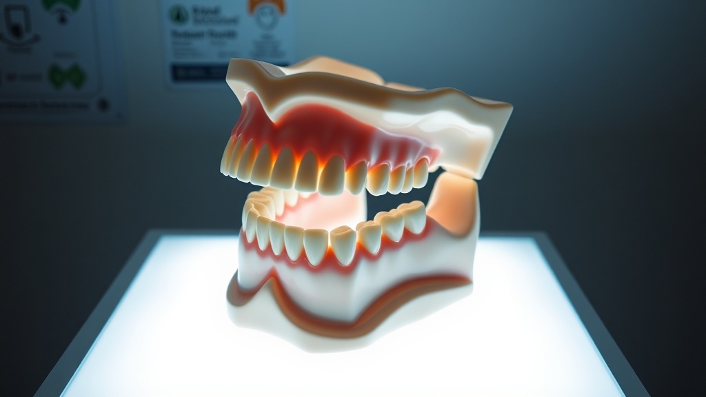
Physical Models and Demonstration Tools
Articulated dental models, extracted tooth specimens showing decay progression, and cross-section models of implant systems provide a tactile, three-dimensional complement to screen-based education. They’re particularly effective for kinesthetic learners and for older patients less comfortable with screen interactions. Handing a patient a model of an implant system and letting them hold it while you explain the procedure creates an engagement that passive viewing cannot replicate.
How Do You Integrate Visual Education Into an Appointment Without Adding Time?
The most common objection to expanding visual education is time: “I don’t have time to add presentations to every appointment.” The reframe is that visual education, when integrated correctly, doesn’t add time — it redirects it. The 3 minutes spent explaining a crown verbally can be replaced by a 90-second animation plus a 90-second conversation that results in better comprehension and faster case acceptance. The net time impact is neutral; the case acceptance impact is positive.
The integration workflow that adds the least time:
- Intraoral camera capture during exam (standard procedure, no additional time): Images are already taken as part of the clinical workflow. The only change is displaying them on a patient-facing screen and narrating findings aloud.
- Radiograph review with patient present (1–2 minutes): Rather than reviewing X-rays alone and then summarizing findings, review them with the patient present and verbalize what you see.
- Procedure-specific animation queued for relevant cases (90 seconds): For any case requiring more than a filling — root canal, implant, periodontal procedure — play the relevant animation while the patient is in the chair. This replaces, not augments, the verbal procedure description.
How Does Visual Education Affect the Patient’s Emotional Experience?
Visual education serves a psychological function beyond information transfer. Patients who understand what is happening in their mouth — who can see the problem and follow the proposed solution — feel more in control of their dental health. That sense of control is a primary driver of reduced dental anxiety, higher treatment acceptance, and greater patient satisfaction.
Research on patient empowerment in healthcare consistently finds that patients who are informed and visually engaged in their own diagnosis make better follow-through decisions, report higher satisfaction with their care, and are more likely to maintain the recall compliance that drives long-term practice retention. Visual education, in this framing, is not just a sales tool — it’s a patient well-being intervention.
For the broader context of how patient education integrates with satisfaction and retention, see our guide to how to improve patient satisfaction in your dental practice. For how visual communication builds the trust that drives referrals, see our pillar guide on building patient trust and relationships.
What Is the Implementation Sequence for Visual Patient Education?
For practices starting from minimal visual education, this sequence builds capacity without overwhelming the team:
Month 1: Implement patient-facing display of existing intraoral camera images during exam. Train team to verbalize findings while images are visible. This costs nothing if you already have an intraoral camera and requires only a positioning change for the monitor.
Month 2: Add patient-facing radiograph review to every new patient exam and any patient with new clinical findings. Annotate digitally where software allows.
Month 3: Add procedure-specific animations for the top 3 procedure types by volume (typically: root canals, crowns, implants). Queue the animations as part of the treatment presentation workflow.
Month 4–6: Implement smile simulation for cosmetic consultations. Build a before-and-after photo library. Establish a patient consent and photo-sharing workflow for social media use.
Measure case acceptance rate at baseline and at 90-day intervals. Most practices see measurable improvement in case acceptance within the first 60 days of consistent intraoral camera use with patient-facing display.
Last Updated: March 2026
For related reading, see our guide on visual case presentation tools that boost treatment acceptance.
Sources
- Journal of Dental Education. “Visual versus verbal patient education in dentistry: effects on treatment plan recall and acceptance.” JDE, 2019.
- BMC Oral Health. “Video-based patient education in dental settings: a systematic review.” BMC Oral Health, 2021.
For more resources on this topic, see our complete guide to patient trust and experience.